Detail Information of Protein
Basic Information:
| Symbol | Hspa9 |
| Synonyms | Grp75; Hsp74; Hspa9a |
| Species | Mouse |
| Entrez ID | 15526 |
| Uniprot ID | P38647 |
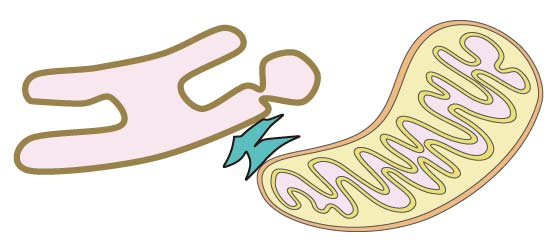
| Membrane Contact Site |
ER-MT; MT-ER
|
| Location (from literature) | MT |
| Cell line/Tissue | Mouse livers; MEFs; Mouse testes; Mouse brains and retinas; RGCs; HRECs; C57BL6 mice |
| Experimental Method | Low throughput experimental methods |
| Protein Sequence |
Complex Information:
| Complex ID | Subunit of complex | Subcellular location | Species | More |
| CMCS00107 | Itpr3; Hspa9; Vdac1; Park7 | ER-MT; MT-ER | Mouse | more | CMCS00161 | Tgm2; Vdac1; Itpr1; Hspa9 | ER-MT; MT-ER | Mouse | more | CMCS00175 | Vdac1; Itpr1; Hspa9 | ER-MT; MT-ER | Mouse | more | CMCS00266 | Itpr3; Hspa9; Vdac1; Pdk4 | ER-MT; MT-ER | Mouse | more |
Expression Overview of Hspa9:
Homology Information of Hspa9:
| Uniprot ID | P38647 |
| EggNOG | KOG0102 |
| HOGENOM | CLU_005965_2_1_1 |
| OrthoDB | 143at2759 |
| TreeFam | TF105046 |
| GeneTree | ENSGT00920000149123 |
References:
| Pubmed ID | 30590033 |
| DOI | 10.1016/j.celrep.2018.11.094 |
| Description | GRP75 localizes in Mitochondrion-associated membranes (MAMs) and acts as a bridging molecule between the two organelles by assembling the IP3R-GRP75-VDAC complex, which is involved in the transport of Ca2+from the endoplasmic reticulum (ER) to Mitochondrion. |
| Description of experimental evidence | The protein was validated by western blot analysis, Co-IP and PLA in mouse livers and MEFs. |
| More related results |
| Pubmed ID | 29785746 |
| DOI | 10.1002/pmic.201700478 |
| Description | By Andromeda search engine against the decoy UniProt-mouse database and Swiss-Prot human database, which yielded 2,808 proteins from mouse testes (mT),2,478 proteins from mouse brain (mB) and 2,155 proteins from human testes (hT). |
| Description of experimental evidence | The protein was validated by TEM and LC -MS/MS analyses in mouse testes. |
| More related results |
| Pubmed ID | 28522876 |
| DOI | 10.1038/s41598-017-02213-1 |
| Description | Using this method, 1313 non-redundant proteins were identified in the MAM. |
| Description of experimental evidence | The protein was validated by proteomic analysis, long gradient nano-reverse-phase liquid chromatography, mass spectrometry and bioinformatics analysis in RGCs, HRECs, mouse brains and retinas. |
| More related results |
| Pubmed ID | 27272971 |
| DOI | 10.1038/srep27351 |
| Description | In this work we describe for the first time the protein composition of highly purified hepatic MAM fractions and demonstrate that CAV1 is a MAM-resident protein. |
| Description of experimental evidence | The protein was validated by stable isobaric labeling and LC-MS/MS in mouse livers. |
| More related results |
| Pubmed ID | 31767755 |
| DOI | 10.1073/pnas.1906565116 |
| Description | DJ-1 regulates the integrity and function of ER-mitochondria association through interaction with IP3R3-GRP75-VDAC1. |
| Description of experimental evidence | The protein was validated by western blotting, electron microscopy, co-immunoprecipitation assays, PLA, Blue-Native and SDS-PAGE 2D separation in C57BL6 mice, and suggests that impaired ER-mitochondria association could contribute to the pathogenesis of PD. |
| More related results |
| Pubmed ID | 24947355 |
| DOI | 10.2337/db13-1751 |
| Description | Mitochondria-associated endoplasmic reticulum (ER) membranes (MAMs) are functional domains between both organelles involved in Ca2+ exchange, through the voltage-dependent anion channel (VDAC)-1/glucose-regulated protein 75 (Grp75)/inositol 1,4,5-triphosphate receptor (IP3R)-1 complex, and regulating energy metabolism. |
| Description of experimental evidence | The protein was validated by immunofluorescence, duolink II in situ PLA and western blot in mouse livers, which regulates energy metabolism. |
| More related results |
| Pubmed ID | 30523025 |
| DOI | 10.2337/db18-0363 |
| Description | Here, we demonstrate that PDK4 interacts with and stabilizes the IP3R1-GRP75-VDAC1 complex at the MAM interface. |
| Description of experimental evidence | The protein was validated by in situ proximity ligation assay, immunofluorescence analysis, coimmunoprecipitation, transmission electron microscopy and immunoblotting in C2C12 myoblasts, which augments ER–Mitochondria contact to dampen skeletal muscle insulin signaling during obesity. |
| More related results |