Detail Information of Protein
Basic Information:
| Symbol | STARD3 |
| Synonyms | CAB1; MLN64 |
| Species | Human |
| Entrez ID | 10948 |
| Uniprot ID | Q14849 |
| Membrane Contact Site |
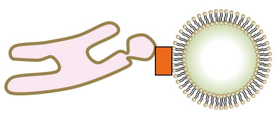
ER-Endosome; Endosome-ER
|
| Location (from literature) | Endosome |
| Cell line/Tissue | HeLa cells; HEK293T cells |
| Experimental Method | Low throughput experimental methods |
| Protein Sequence |
Complex Information:
| Complex ID | Subunit of complex | Subcellular location | Species | More |
| CMCS00014 | MOSPD2; STARD3 | ER-Endosome; Endosome-ER | Human | more | CMCS00023 | VAPA; VAPB; STARD3 | ER-Endosome; Endosome-ER | Human | more |
Expression Overview of STARD3:
Homology Information of STARD3:
| Uniprot ID | Q14849 |
| EggNOG | KOG3845 |
| HOGENOM | CLU_033480_0_0_1 |
| OrthoDB | 3740290at2759 |
| TreeFam | TF313869 |
| GeneTree | ENSGT00940000159051 |
References:
| Pubmed ID | 24105263 |
| DOI | 10.1242/jcs.139295 |
| Description | STARD3 or STARD3NL and VAP form a novel molecular tether between late endosomes and the ER. |
| Description of experimental evidence | The protein was validated by immunoprecipitation, electron microscopy, immunofluorescence, in situ proximity ligation assay, mSDS-PAGE and western blot analysis in HeLa cells. |
| More related results |
| Pubmed ID | 28377464 |
| DOI | 10.15252/embj.201695917 |
| Description | Corroborating this, in vitro reconstitution assays indicated that STARD3 and its ER-anchored partner, Vesicle-associated membrane protein-associated protein (VAP), assemble into a machine that allows a highly efficient transport of cholesterol within membrane contacts. |
| Description of experimental evidence | The protein was validated by immunofluorescence, colocalization analysis, filipin staining, GFP-D4, filipin co-staining, flotation experiments, electron microscopy and stereology in Hela cells, and the complex allows a highly efficient transport of cholesterol within membrane contacts. |
| More related results |
| Pubmed ID | 29858488 |
| DOI | 10.15252/embr.201745453 |
| Description | Consequently, MOSPD2 and these organelle‐bound proteins mediate the formation of contact sites between the ER and endosomes, mitochondria, or Golgi.; ll these proteins, by binding VAP proteins, are known to build contact sites between the ER and endosomes (STARD3, STARD3NL, ORP1L), mitochondria (PTPIP51), and Golgi (STARD11). |
| Description of experimental evidence | The protein was validated by immunofluorescence, colocalization analysis, SDS–PAGE, western blot, coomassie blue staining, pull‐down assays, GFP‐Trap, mass spectrometry analysis, electron microscopy and immunoprecipitation in HeLa cells and HEK293T cells. |
| More related results |